MSU anthropologist member of $1.4 million NIH grant team to develop image library for facial features requiring surgery
December 7, 2021 - Katie Nicpon

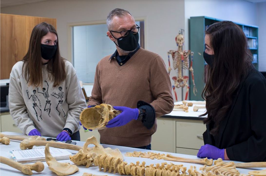
Photo: MSU Assistant Professor of Anthropology Joseph T. Hefner (middle) working with graduate students in the new MSU Forensic Anthropology Laboratory. The skeleton was donated for educational purposes. Photo credit: Jackie Belden Hawthorne.
The National Institute of Dental & Craniofacial Research of the National Institutes of Health (NIH) awarded Joseph T. Hefner, Ph.D., and colleagues at the University of Kentucky a five-year, $1.4 grant to develop a standardized graphic library to assist clinicians and biomedical researchers to communicate anatomical concepts with their patients and their families. Hefner is an MSU Department of Anthropology assistant professor and CO-PI on the grant team led by Melissa Clarkson, Ph.D., assistant professor of biomedical informatics from the University of Kentucky. The project, titled, “Developing standardized graphic libraries for anatomy: A focus on human craniofacial anatomy and phenotypes,” began Summer of 2021.
“The focus is on creating standards for clinicians and surgeons who meet with patients and their family members and engage them about the type of craniofacial anomaly they have and the necessary reconstructive surgery,” Hefner said.
Craniofacial anomalies are irregular facial features, such as cleft palate or cleft lip, and might require surgery. According to the Centers for Disease Control and Prevention , about one in every 1,600 babies is born with cleft lip and cleft palate in the United States.
“We are generating illustrations that can be used to guide family members through the process of understanding anomalies like cleft lip or cleft palate,” Hefner said. “We have the clinical language which is very complex, but with the addition of line drawings, clinicians can talk to family members in a more easily understood way and using tools to help the family visualize how the reconstruction will proceed.”
The team is led by Clarkson who specializes in addressing the gap between complex information and the people who need to understand it. The team members represent an interdisciplinary approach that includes a plastic surgeon, an odontologist, a traditional human anatomist, and Hefner, a biological anthropologist. Hefner’s knowledge and research on global human craniofacial variation is a key component of the project from a perspective of equity. The earliest models for the current clinical language and images of craniofacial anomalies are based upon American white populations.
“These models really miss some of the nuances of craniofacial morphology here in the United States, and my job is to provide data and an expertise on human variation to more accurately capture craniofacial variation,” Hefner said.
In other words, Hefner’s contribution will help to shape clinical language and imagery that reflect the diverse people in the United States.
“For example, ‘wide mouth’ is defined clinically as ‘the distance between the corners of the mouth greater than two standard deviations above the mean,’ - but what does that look like in a living individual?” Clarkson explained. “Drawing that phenotype [set of observable characteristics] will depend on population-level data, and that data should reflect different ages and populations. Dr. Hefner will help us to understand population-level differences in phenotypes and how to incorporate craniometric [ the scientific measurement of skulls] and macromorphoscopic [soft tissue differences] datasets into our work.”
Additionally, their research will serve as standardized visual representations for information systems and software applications. For example, their work will include developing prototypes for web-based tools such as the Human Phenotype Ontology .
This project is a new application of Hefner’s research. Much of his previous work has involved forensic anthropology and working with medical and legal death investigations.
“The exciting thing for me is that I’ve never done anything like it,” Hefner said. “I can use my love of human craniofacial morphology for a far-reaching, great cause. We’re dealing with people who have craniofacial anomalies, which is very common here in the United States, to provide them and their families and clinicians a common visual language, improving discussions between patients and doctors.”

